
AF. SEM of lingual mucosa of C. niloticus. A and B. Showing mucous
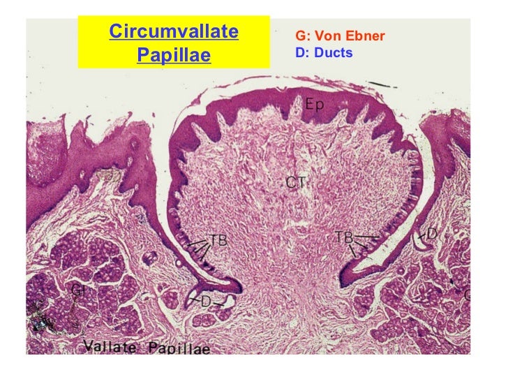
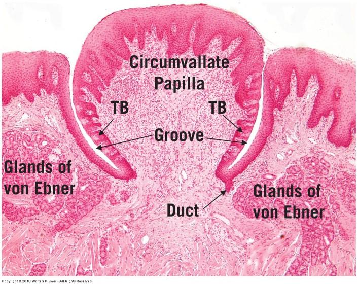
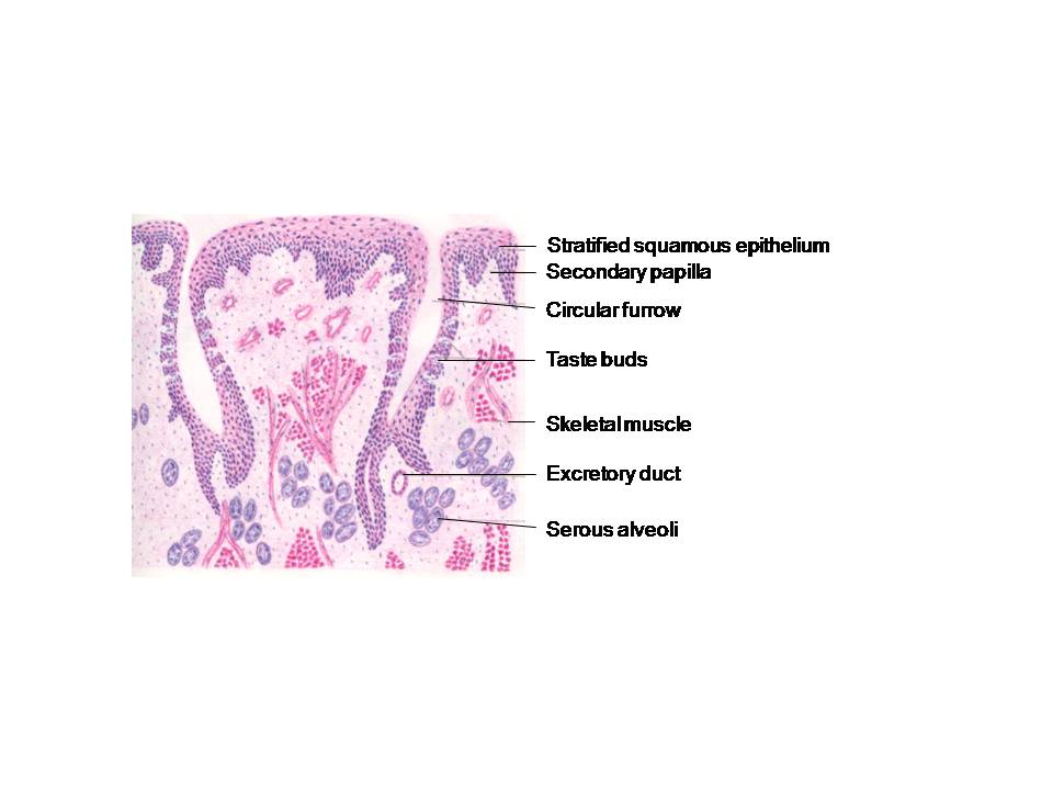
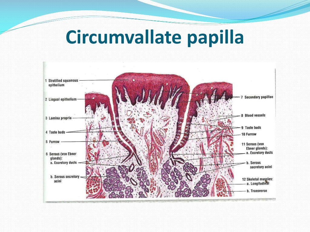
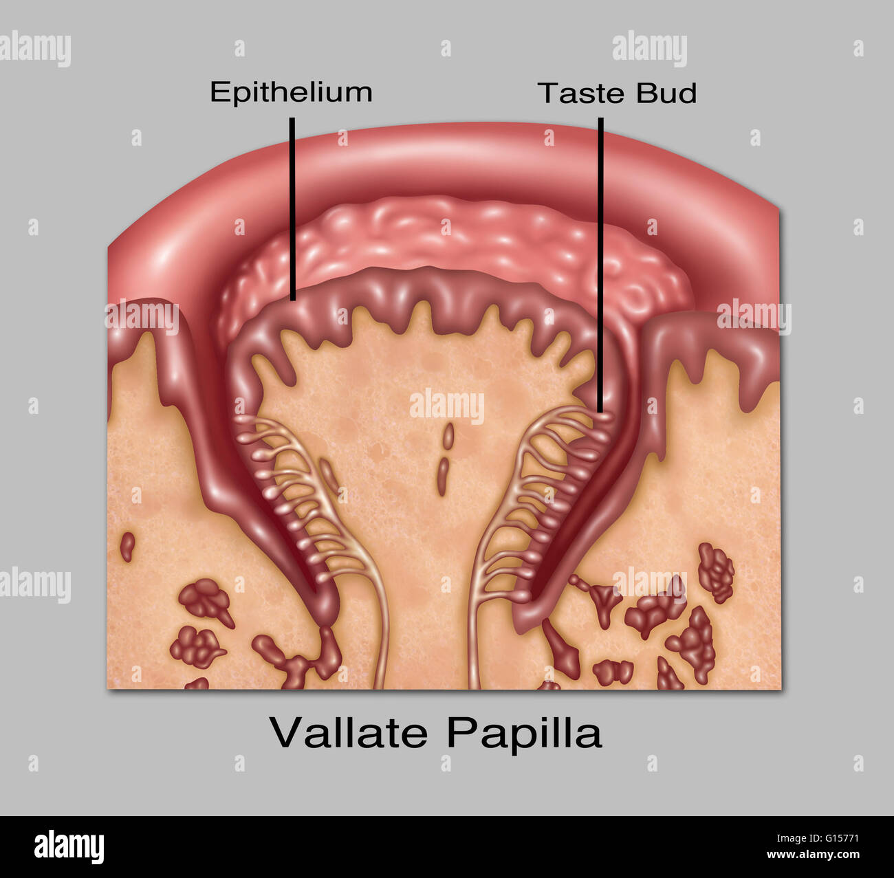
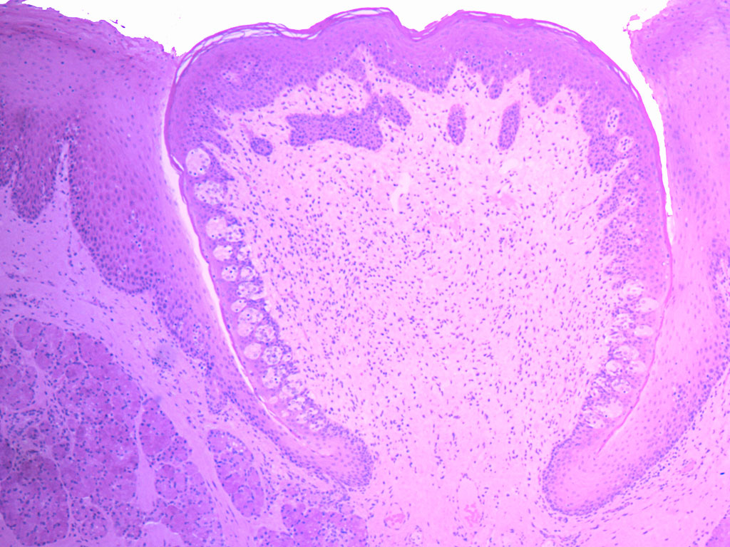
Tongue: Circumvallate papillae. Each circumvallate papilla is surrounded by a deep, circular sulcus into which the serous glands of von Ebner empty. The connection between the duct of the glands and the sulcus is visible. The secretory cells of the glands contain apical eosinophilic granules; their watery secretions cleanse the surfaces of the.
Lip & tongue
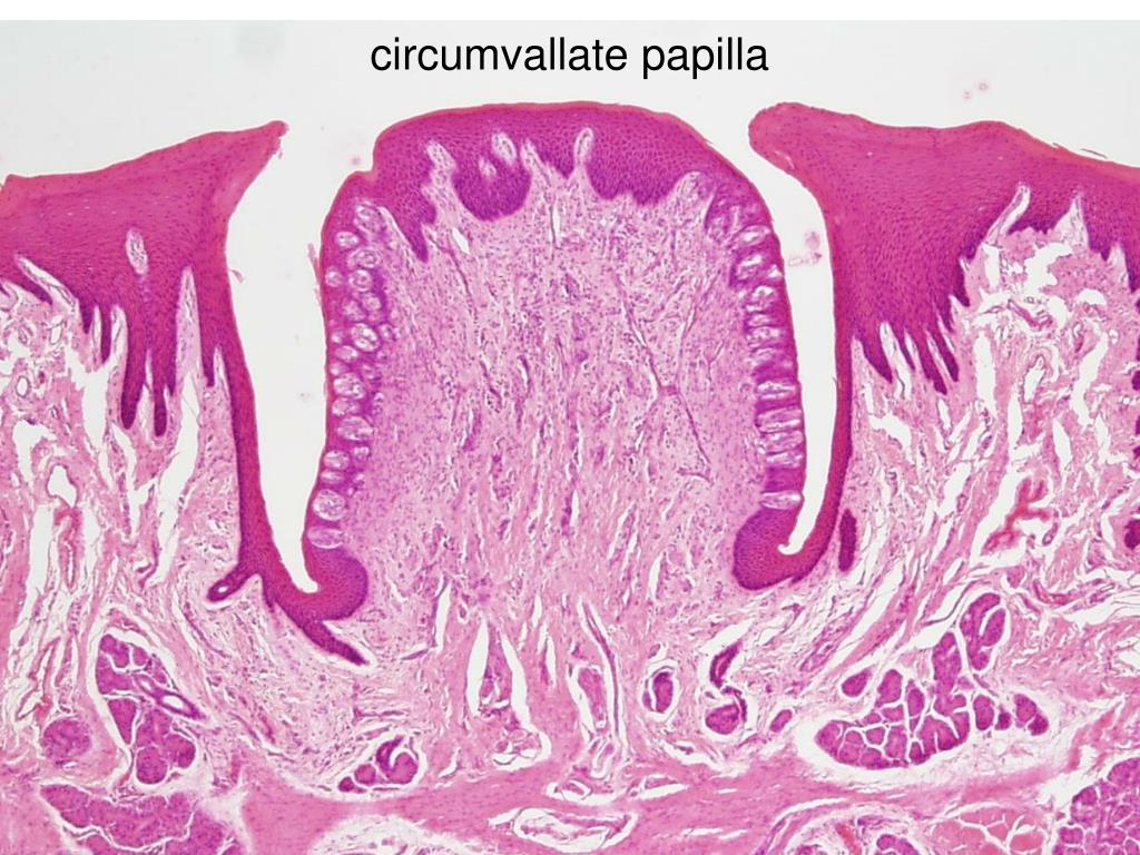
Vallate papillae: These papillae lie in a V-shaped row immediately anterior to the terminal sulcus, which divides the dorsum of the tongue into its anterior two-thirds and a posterior third.. Vallate papillae are round in shape. Their apex is coated with stratified squamous epithelium. About 50% of all taste buds are found in the circumvallate papillae.

[PDF] Histological study of tongue in insectivore bat (Rhinopoma
The number of circumvallate papillae among the rodents is widely different. The one large papilla surrounded by roll is located on the posterior part on the medial line of tongue not only in white laboratory rat but also in mouse and bank vole . Flying squirrel, shrew, and American beavers have three circumvallate papillae [10, 15, 18-21].
PPT Taste PowerPoint Presentation, free download ID268846
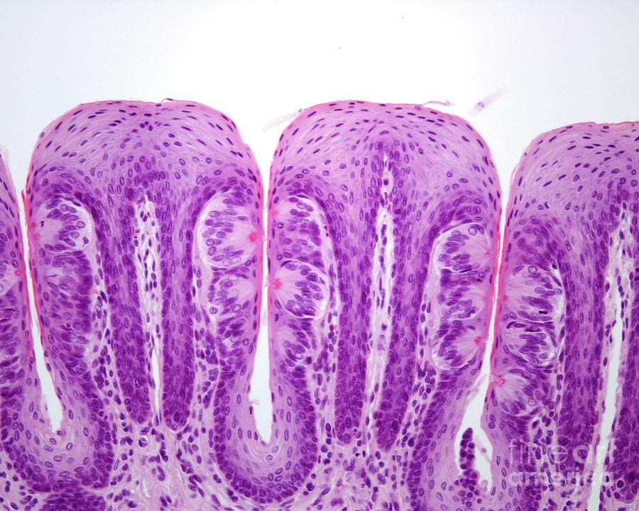
Tongue: Filiform papillae. The dorsal and lateral surfaces of the tongue are covered by specialized mucosa forming papillae: filiform, fungiform, circumvallate, and foliate. The tapering filiform papillae seen here are the most numerous type, covering most of the anterior two thirds of the tongue. Each papilla is about two to three millimeters.
Print Vertebrate Histology Exam 4 flashcards Easy Notecards
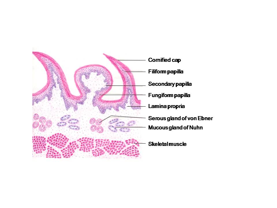
These papillae are mechanical and do not contain taste buds. Circumvallate Papillae - eight to twelve dome-shaped structures in a V-shaped row in front of sulcus terminalis. The largest and least numerous papillae found on the tongue. Furrow - each papillae is surrounded by a moat (or sulcus) that receives saliva from minor salivary glands.
circumvallatepapillaslidelabelledhistology SchoolWorkHelper
Tongue: Circumvallate papillae. Eight to twelve circumvallate papillae are located along the sulcus terminalis, separating the anterior from the posterior portion of the tongue. Each papilla is surrounded by a deep sulcus that receives ducts of the serous glands of von Ebner. The connective tissue core is covered on its lateral surfaces with.
HISTOLOGY DIAGRAMS February 2016
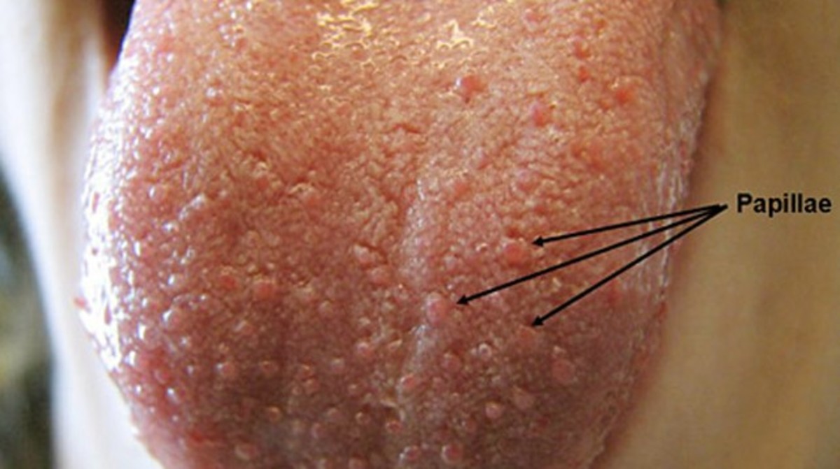
Lingual papillae ( SG: papilla) are small structures on the upper surface of the tongue that give it its characteristic rough texture. The four types of papillae on the human tongue have different structures and are accordingly classified as circumvallate (or vallate), fungiform, filiform, and foliate. All except the filiform papillae are.

Oral Histology Circumvallate Papillae Adult Tongue at Rs 800/piece in
Circumvallate papillae (also known as vallate papillae) are organized linearly, as a set of four to six large papillae anterior to each limb of the sulcus terminalis (total = 8-12 papillae).. Ross MH, Pawlina W. Histology: a text and atlas with correlated cell and molecular biology. 8th ed. Amsterdam: Wolters Kluwer Health; 2017. Google.
Enlarged Papillae Pictures, Causes and Treatment HubPages
Circumvallate (Vallate) Papillae. Circumvallate papillae (also known as vallate papillae) are organized linearly, as a set of four to six large papillae anterior to each limb of the sulcus terminalis (total = 8-12 papillae). The characteristic furrow found within the papillae can be appreci-ated. These moats facilitate drainage of serous salivary

Oral Histology Circumvallate Papillae Fetal at Rs 800/piece in Noida
These papillae are less readily observed in adults, because of slight keratinization of the epithelium. Slide 117 and especially slide 117N contain examples of circumvallate papillae View Image. These are large circular papillae surrounded by a deep trench. The covering epithelium is non-keratinized.
PPT Histology of Tongue, Liver & Pancreas PowerPoint Presentation
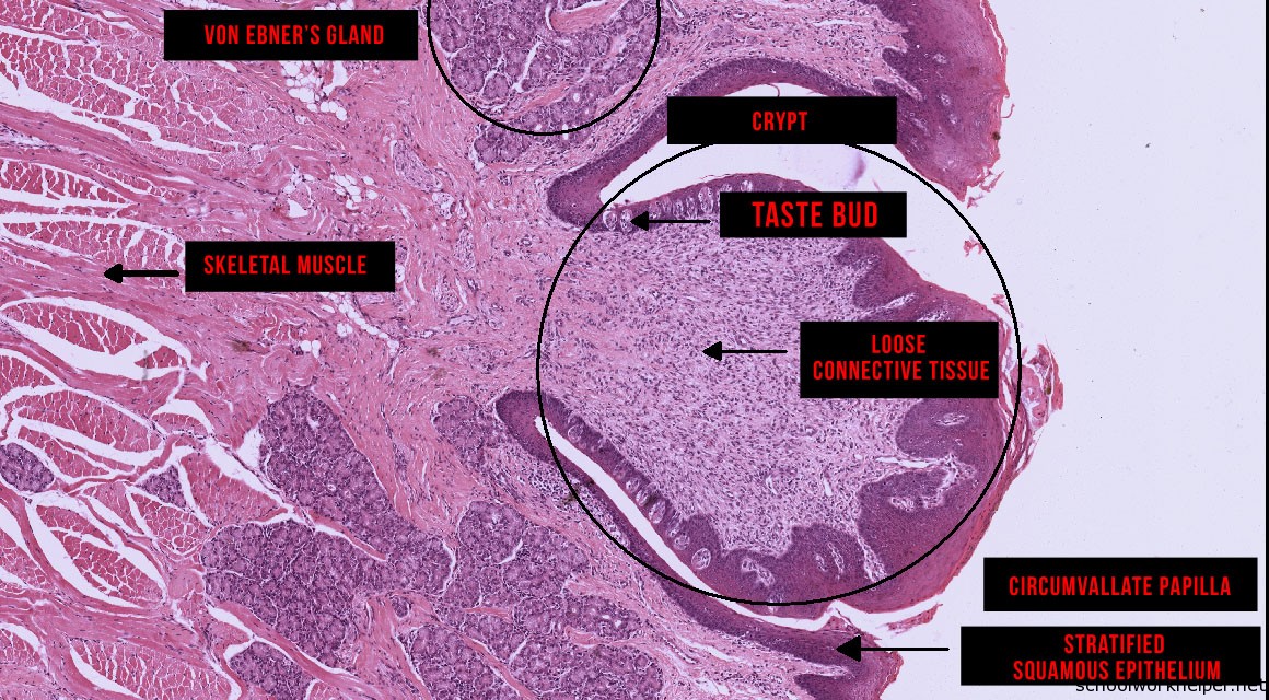
Histology @ Yale. Slide List. Circumvallate Papilla. Circumvallate Papilla Circumvallate papillae are the largest papillae on the tongue. They are covered with a stratified epithelium and the walls contain numerous taste buds.
Circumvallate papillae hires stock photography and images Alamy
Tongue: Circumvallate papillae. Along the lateral edges of the circumvallate papillae are oval taste buds, which access the oral cavity through a taste pore. Taste buds are composed of supportive cells and neuroepithelial cells that are innervated by sensory neurons of the facial nerve (cranial nerve VII). 40x, 400x.
Circumvallate Papilla
These papillae (namely filiform, fungiform, foliate, and circumvallate) play different roles in perception of general and special sensory stimuli. The lingual papillae (except the filiform type) and other areas of the lingual mucosa, have taste buds scattered across their surfaces. These are specialized collections of taste cells that.
Foliate Papillae With Taste Buds Photograph by Jose Calvo / Science
General afferent impulses from the circumvallate papillae, along with the posterior third of the tongue are carried by fibers of the glossopharyngeal nerve (CN IX). Lingual nerve Nervus lingualis 1/3.. Junqueira's Basic Histology. 13th ed., Mcgraw-Hill, 2013. Moore, Keith L et al. The Developing Human. 9th ed., Elsevier-Saunders, 2013.

Enlarged Circumvallate Papillae Pictures, Causes, Treatment, Cancer
In the vallate and circumvallate papillae of the animal's tongue, you will find some special histological features - It is a sunken inverted cone-shaped papilla with a flat top that lines with the stratified squamous epithelium, Presence of numerous taste buds on the lateral wall of the circumvallate papillae,
HISTOLOGY DIAGRAMS Special histology specific points
Taste buds: barrel shaped, lightly staining, intramucosal sensory receptors present in large numbers on circumvallate papillae and in lesser numbers elsewhere. Intraepithelial nonkeratinocytes: melanocytes (basal), Merkel cells (basal), Langerhans cells (suprabasal) and lymphocytes occur in oral mucosa. Tonsillectomy specimens frequently.